Send us your feedback
Here you can send us feedback on the Maxess-website. Please describe the problem or what’s missing in a clear way, and on what page you found the issue. Thank you so much for your help!
High speed X-ray radiography allows for the measurement of powder-based drugs inside nasal inhalers
The biopharmaceutical company Iconovo AB in Lund together with MAX IV imaging experts carried out high speed Synchrotron X-ray radiography at the ID19 beamline at ESRF (European Synchrotron Radiation Facility, Grenoble, France) for studying the behaviour of powder-based drugs inside nasal inhalers. These experiments also confirmed that Iconovo’s computational flow simulations of the behaviour of the powder-based drugs inside the nasal inhalers during intake were accurate.
Nasal inhalers as a drug and vaccine delivery system
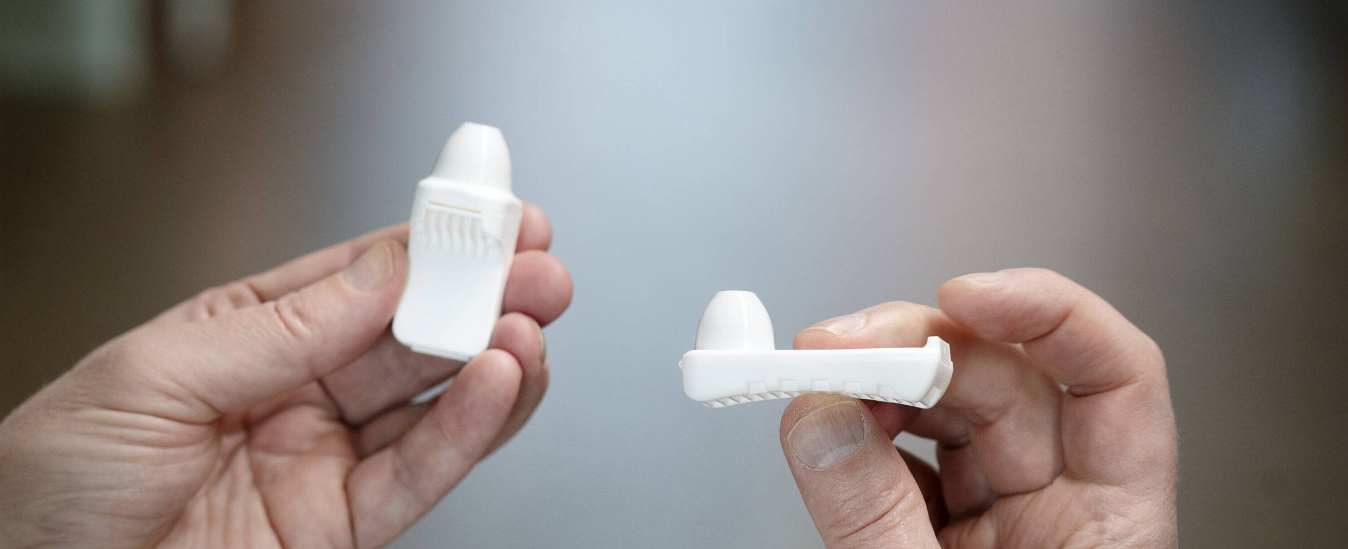
Nasal inhalers are used as a drug delivery system for powder-based drugs to treat conditions such as asthma or to administer vaccines. The product design of nasal inhalers determines the dynamic flow behaviour of the powder-based drugs and hence their efficiency. This is usually determined via computational flow simulations but to confirm their accuracy, and find irregularities or unexpected flow patterns and behaviours, practical experiments are required. Iconovo wanted to examine this powder movement and behaviour during inhalation for their ICOone nasal inhaler using such experiments. To do so, they participated in a collaborative project involving expertise and equipment from Lund University (LU), MAX IV and ESRF with funding from LEAPS-Innov, all mediated via the MAXESS project.
Optimisation experiments using lab-based radiography are required before moving to the synchrotron
As a starting point, initial lab-based X-ray radiography experiments were conducted using an Rx Solutions scanner (Chavanod, France) at the 4D imaging Lab at the Division of Solid Mechanics, Faculty of Engineering, LU, to carry out pre-tests, optimise the experimental conditions and the sample environment (Figure 1A). A trigger box connected to a vacuum pump and the inhaler mimicked the intake of a powder-based drug via the nose cavity, at 5 frames per second (Figure 1B & Figure 2), which also motivated the need for a higher number of frames per second. It also provided valuable information for the beamline staff at ESRF about setting up the beamline with respect to the camera settings (FOV-field of view), exposure time (for optimal contrast) and beam energy required.
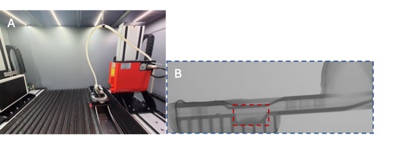
Figure 1. (A) Set up at the Rx scanner at the 4D Imaging Lab, LU, and (B) resulting radiograph, with the entire FOV (58 mm in width) in blue, and the selected region of interest for the final experiment at ESRF.

Figure 2. Radiograph of the powder in the inhaler with the RX scanner from the highlighted region from Figure 1B. Frames per second (during acquisition): 5, FOV: 30 mm in width. Please note that the contrast is sufficient to resolve clusters of powder, although the resolution is too low to resolve single powders.
High-speed synchrotron X-ray radiography is a non-destructive time-resolved 2D technique capable of studying dynamic flows in real time
In addition to the lab-based preliminary experiments, a few improvements on the set up were added including a 3D printed nose cavity (Figure 3). The ID19 beamline at ESRF allows for very fast radiography images, with measurements of 10,000 frames per second, allowing for imaging and measurements in real-time.
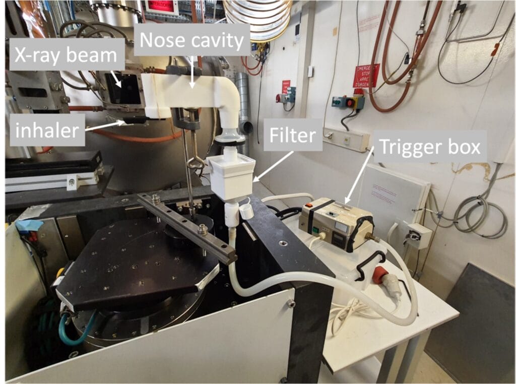
Figure 3. Final experimental set up at the ID19 beamline at ESRF, including a standardized and representative nose cavity.
Whereas the initial experiments could only see the location of the powder at the start and end, the synchrotron experiments allowed visualisation and measurement of the entire process of powder movement during inhalation (Figure 4 and 5).
The results from ESRF confirmed that Iconovo’s computational flow simulations of the behaviour of the powder-based drugs inside the nasal inhalers during intake were accurate. No other technique has so far been able to confirm the simulations up until now.
The project MAXESS SmiLe’s: SMEs to LRSI allowed Iconovo access to expertise, training and facilities which are often difficult for SMEs to do alone, equipping them with the necessary knowledge to do future experiments unaided. Not only did this experiment provide valuable input into final product design but it also equipped Iconovo with a new method for their inhalation research.
Lab scale experiments in radiography are vital before going to synchrotrons and provide important information with regards to optimizing the set up.
Emanuel Larsson, tomography expert, MAX IV and Lund University
By using the synchrotron we have gained new knowledge of how the powder behaves in our devices, which gives us an opportunity to optimise the design and increase the performance of our inhalers
Simon Karlsson, Mechanical Engineer, Iconovo AB
Contact Partners
Case Details
Iconovo AB
Iconovo ABSmile IncubatorCIPA4D Imaging LabESRF Business Development Office